Translate

Friday, February 20, 2015

Carotid Stenosis (Carotid artery disease)

Carotid stenosis (stenosis = narrowing) is the narrowing of carotid arteries in the neck. Carotid arteries carry oxygen-rich blood to a large part of the brain. Gradual narrowing of these arteries causes decrease blood flow to the brain leading to stroke. There are no community based studies describing the incidence and prevalence of carotid artery disease in India. The increasing prevalence of heart disease, diabetes and other risk factors such as obesity only indicates that the incidence of carotid stenosis in India is on the rise. Hence it is important for family doctors and people to be aware of this condition and seek appropriate treatment.

What is carotid stenosis?
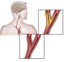
Progressive narrowing of carotid arteries in the neck occurs due to the formation of plaque consisting of fat, cholesterol and calcium. Over time, this plaque gradually prevents adequate amount of blood going to the brain. People with carotid stenosis have an increased risk of having a stroke in one of the three ways:

  • The plaque may grow larger and larger and may eventually completely block the carotid artery.
  • Due to decreased blood flow through the carotid arteries, there is an increased risk of forming blood clots in the carotid artery. These blood clots may go into the brain and block one of the major arteries.
  • The plaque may break and block one of the arteries in the brain.
What are the risk factors for carotid stenosis?
Carotid artery stenosis.png
  • Age
  • Smoking
  • High blood pressure
  • High 'bad' Cholesterol (LDL)
  • Diabetes
  • Obesity
  • Family history of heart disease and stroke
What are the symptoms?
Unfortunately, many people with carotid artery disease do not have symptoms until the late stage and, in many patients stroke may be the first indication of carotid stenosis. Transient ischemic attack (TIA) or 'Warning Stroke' may be another indication of underlying carotid artery disease. Common symptoms of TIA include

  • Sudden numbness, tingling, weakness, or loss of movement in your face, arm, or leg, especially on only one side of your body. 
  • Sudden vision changes. A special type of TIA is amaurosis fugaxTransient blindness in one eye occurs because debris from a narrowed carotid artery clogs the artery (ophthalmic artery) that supplies blood to the retina of the eye. 
  • Sudden trouble speaking. 
  • Sudden confusion or trouble understanding simple statements. 
  • Sudden problems with walking or balance.
  • How is carotid stenosis diagnosed?
    Every doctor should be aware of this condition and should refer the patient to the appropriate specialist when in doubt. A Stroke Neurologist is the best person to diagnose and  manage carotid artery disease initially. Other than a thorough clinical examination, some of the common tests used to diagnose carotid stenosis and plan treatment are

    • Carotid doppler ultrasound
    • CT angiography
    • MR angiography
    • Digital Subtraction Angiography
    How is carotid stenosis treated?
    • Lifestyle modification slows the growth of the plaque. This is the most important step in treatment of carotid stenosis
    • Depending upon the associated medical conditions, your doctor may prescribe blood thinners such as aspirin, clopidogrel and warfarin. Statins have a major role to play in the medical management of carotid artery disease. High blood pressure, diabetes and heart disease, if present, should be treated appropritely
    • Carotid Endarterectomy: In selected patients, surgery may be required to remove the plaque. Surgery involves making an incision in the neck, opening the carotid artery, removing the plaque and sewing the artery back to restore blood flow to the brain
                                               
    • Carotid angioplasty and stenting: In patients with severe uncontrolled heart disease in whom surgery may carry a high risk, opening the artery from inside and placing a stent by inserting a catheter through the groin may be an option.

    Friday, February 13, 2015

    Lifestyle Changes to Prevent Stroke

    The annual incidence of stroke in the country has increased from 13 per 100,000 in 1969 to 145 per 100,000 in 2006. The costs of acute care and managing these patients in long term are astounding. In another study, the overall DALYs (Disability Adjusted Life Years) lost due to stroke were 795.57 per 100,000 person years. This means that if 100,000 people were to live for one year, about 800 people will be disabled due to stroke and will not be able to go to work.

    As individuals, we should be aware of the healthy ways of living to prevent a stroke. In this article, I will discuss some of the lifestyle changes that we can adopt in order to decrease the risk of having a stroke and heart attack.

    How to make lifestyle healthier?
    • Do not smoke. Smoking is one of the strongest predisposing factors for stroke and heart attack. Passive smoking is harmful as well. Ideally, it's best to not start smoking in the first place, but if you're a current smoker, no matter what your age, quitting can be extremely beneficial. For example, if you quit smoking before the age of 50, you cut your risk of dying in the next 15 years in half compared to those who keep smoking.
    • Eat healthy. Eat foods low in saturated fat, trans fat, cholesterol, sodium and added sugars. Some of the nutrients sources for vegetarians are
      • Proteinschickpeas, lentils, green peas, soybeans and kidney beans, beans, nuts, nut butters, peas, and soy products (tofu). Milk products and eggs are also good protein sources for lacto-ovo vegetarians. Calcium-fortified nondairy substitutes, such as soy milk and soy yogurt, are also excellent sources of protein. A cup of milk or soy milk provides about 8 grams, and 1 cup of low-fat yogurt contains about 13 grams of dietary protein. Paneer, a fresh cheese common in Indian cuisine, contains about 7 grams of protein per ounce
      • Ironiron-fortified breakfast cereals, spinach, kidney beans, black-eyed peas, lentils, turnip greens, whole wheat chappati, peas, and some dried fruits (dried apricots, raisins)
      • Calciumcalcium-fortified milk, calcium-fortified breakfast cereals and orange juice, tofu made with calcium sulfate, and some dark-green leafy vegetables (collard greens, turnip greens, bok choy, mustard greens). Milk products are excellent calcium sources for lacto vegetarians.
      • Zincbeans (white beans, kidney beans, and chickpeas), zinc-fortified breakfast cereals, wheat germ, and pumpkin seeds. Milk products are a zinc source for lacto vegetarians.
      • Vitamin B12 - milk products, eggs, and foods that have been fortified with vitamin B12. These include breakfast cereals, milk and nutritional yeast.
      • Avoid foods like egg yolks, fatty meats, butter and cream, which are high in fat and cholesterol.
      • Eat moderate amounts of food and cut down on saturated fat, trans fat, sugar and salt.
      • Bake, broil, roast and boil foods instead of frying.
      • Read nutrition labels on packaged meals. Many are very high in sodium.
      • Limit alcohol to one drink a day for women; two drinks per day for men.
      • Eat more fruit, vegetables, whole-grains, dried peas and beans, pasta, fish, poultry and lean meats.
      • Ask your doctor, nurse or a nutritionist or dietician for help.
    • Be physically active
      • Look for small chances to walk or exercise. Take the stairs instead of the elevator
      • Brisk walking or running for at least 2-3 hours a week is good for health
      • Consult your doctor before starting any exercise if you have a medical condition
    • Get regular blood pressure checks. Don't be afraid of high blood pressure. Work with your doctor to control the blood pressure
    • Maintain a healthy weight. A basal metabolic index (BMI) of less than 25 is acceptable
    • Take your medicines as directed by the doctor
    • Reduce the stress level in your daily life. Meditation helps
    • Have regular medical check-ups. Do not ignore warning signs

    Friday, February 6, 2015

    'Transient Ischemic Attack' or 'Mini-Stroke'

    A 'Transient Ischemic Attack' (TIA) or 'Mini-stroke' is a condition in which the person experiences the symptoms of a stroke for a few minutes. The symptoms completely resolve within a few minutes. About 1 in 3 persons experiencing TIA will eventually develop a full blown stroke. This is the reason why TIA is also called 'Warning Stroke'. It also gives the person time to act and prevent permanent stroke.

    TIA occurs because the blood flow to a part of the brain stops temporarily due to a clot. When the body dissolves the clot, blood flow is restored and the symptoms resolve. Decreased blood flow due to narrowing of an artery supplying a part of the brain may also lead to TIA in situations where the demand increases.

    The common symptoms of TIA are exactly the same as that of stroke.
    • Sudden numbness, tingling, weakness, or loss of movement in your face, arm, or leg, especially on only one side of your body.
    • Sudden vision changes. A special type of TIA is amaurosis fugaxTransient blindness in one eye occurs because debris from a narrowed carotid artery clogs the artery (ophthalmic artery) that supplies blood to the retina of the eye.
    • Sudden trouble speaking.
    • Sudden confusion or trouble understanding simple statements.
    • Sudden problems with walking or balance.
    Most symptoms of TIA disappear in a few minutes to hours.

    Risk factors for TIA are the same as that for stroke

    • strong family history of stroke
    • smoking
    • high cholesterol
    • high blood pressure
    • heart disease
    • diabetes
    Tests that your doctor may perform

    • Tests that show pictures of your brain and blood vessels, such as a CT scan, an MRI, a magnetic resonance angiogram (MRA), or an angiogram.
    • A test that uses sound to check your blood flow (Doppler ultrasound).
    • An echocardiogram (echo) to check your heart's shape and its blood flow.
    • An electrocardiogram (EKG, ECG) to measure your heart rhythm.
    • Blood tests, including a complete blood count and a fasting blood test to check for problems that could be causing your symptoms.
    The goal of TIA treatment is to prevent a full blown stroke. Treatment depends on the exact cause of the TIA. In addition to lifestyle changes such as diet, physical activity, limiting alcohol intake, and not smoking, the doctor may recommend medications to treat high blood pressure, high cholesterol, or heart disease. These changes may reduce your risk of further TIA or stroke. There are many medications that help prevent blood clots from forming, thus, reducing the risk of full-blown stroke.
    If a TIA is caused by blockage in the main artery in the neck that supplies blood to the brain, called the carotid artery, surgeries may be required to open the artery, and prevent a stroke. These procedures are known as endarterectomy and stenting.
    If TIAs are appropriately treated on time, one can prevent stroke and major disability. Hence, it is essential for people to be aware of this condition and seek treatment as soon as possible.

    Friday, January 30, 2015

    Moyamoya disease

    Moyamoya disease is a rare disorder of the carotid arteries - the arteries that supply blood to the brain. The term 'Moyamoya' means 'puff of smoke'. It refers to the appearance of arteries on a cerebral angiogram. The condition was first described in Japan, and, since then has been described in other parts of the world.

    What is Moyamoya disease?

    In this condition, the carotid arteries that supply blood to the brain become thickened and narrowed gradually over time. As a result, the brain does not receive enough oxygen-rich blood. To compensate for the reduced supply, the body grows new arteries around the head and brain. These new arteries, also called the 'Moyamoya vessels' try to compensate for the reduced blood flow. However, these new vessels never fully compensate for the reduced blood supply. They also have a propensity to bleed easily.

    Who does Moyamoya disease affect?

    There are no community based studies from India. The prevalence of the disease ranges from 3.2 to 10.5 per 100,000 population. In general, the disease has been found to be more prevalent among Asians and people of Asian origin. The exact cause of this disease is not known yet. About 57% of the affected patients are Asian and 71% are female. Although the disease may be seen in any age group, it is more common in people from 5-15 years and 30-40 years of age. Family history is present in about 10%-15% of the patients.

    How does Moyamoya disease present?

    All the symptoms of Moyamoya disease arise from reduced blood supply to the brain and/or rupture of the 'Moyamoya vessels'. Reduced blood supply may cause stroke and rupture of the unhealthy Moyamoya vessels causes bleeding within the brain. Adults experience hemorrhage more commonly; cerebral ischemic strokes from reduced blood supply are more common in children.

    Children may have weakness or numbness of an arm or leg, hemiparesis, monoparesis, involuntary movements, headaches, dizziness, or seizures. Mental retardation or persistent neurologic deficits may be present.

    Adults may have symptoms and signs similar to those in children, but hemorrhage (bleeding) of sudden onset is more common in adults.

    How is it diagnosed?

    The condition is diagnosed on MRI and cerebral angiography in a patient presenting with stroke or brain bleed.

    Are there any associated conditions?

    Many conditions are seen more commonly with Moyamoya like condition, although the exact cause-effect relationship has not been demonstrated. Some of the common conditions are

    • Radiotherapy to head and neck
    • Down's Syndrome
    • Neurofibromatosis type 1
    • Sickle cell disease
    • Congenital Heart Disease


    Is there treatment?

    Since the exact cause of the disease is unknown, there is no treatment to reverse the narrowing. However, treatment is aimed at preventing stroke by administering aspirin and surgery that improves blood flow to the affected parts of the brain. Surgery is highly successful in preventing stroke and bleeding in future and is the mainstay of treatment.

    Without surgery, the majority of individuals with Moyamoya disease will experience mental decline and multiple strokes because of the progressive narrowing of arteries. 

    Tips for family and caregivers of patients with Moyamoya disease

    Educating and supporting the child and family plays a key role. Not surprisingly, parents are shocked and frightened when they learn their children have had strokes, because they do not realize children can have strokes. Parents should be instructed to inform surgeons and anesthesiologists to avoid hyperventilation. Parents and children need to identify everyday events that may precipitate mini-strokes (also called Transient ischemic attacks). For example, one patient had a stroke while crying or singing a long note during practice. The precipitation of mini-strokes by everyday events is particularly stressful for parents, who may be reluctant to discipline their child for fear of causing a mini-stroke if the child cries. Some sports such as cricket and soccer that lead to hyperventilation, have a high risk of causing mini strokes, and it may not occur to parents to keep their children out of these sportsSchools should be informed about the diagnosis and any restrictions on physical activity. 

    Educating the child and family about seizures is an important part of the care of moyamoya patients. As with other seizure patients, families should be told that brief seizures lasting a few minutes are not thought to be harmful, but medical help should be sought for longer seizures.

    Emotional support and appropriate advice on pre and post operative care of the patient is an important part of treatment to alleviate the fear, anxiety and uncertainity experienced by the family.

    Friday, January 23, 2015

    Venous Stroke

    'Venous Stroke' or 'Cerebral Venous Sinus Thrombosis' is caused by blood clots blocking the veins in the brain. The veins in the brain drain blood from the brain into the heart. When the veins are blocked, the blood cannot drain normally and causes back pressure in the brain leading to all the symptoms. Although venous strokes account for about 1% of all the strokes across all age groups and about 3/4th of all the strokes in people younger than 50 years.

    Causes

    Any predisposing condition that leads to increased tendency for blood clot formation, can cause venous stroke. The stroke occurs across all age groups, from infants , pregnant women, cancer patients, patients with blood disorders to elderly patients. Women tend to be more often affected than men.

    Predisposing conditions

    • Infection
    • Trauma
    • Pregnancy, puerperium and birth control pill use
    • Blood disorders that lead to increased tendency to form blood clots
    • Bowel diseases such as Crohn's disease and Ulcerative colitis
    • Cancer

    Symptoms
    1. Sudden weakness or numbness of face, arm or leg; especially if the numbness is all on one side of the body
    2. Sudden confusion, trouble speaking or understanding
    3. Sudden trouble seeing in one or both eyes
    4. Sudden trouble walking, dizziness, loss of balance or coordination
    5. Sudden severe headache with no known cause
    6. New onset seizures

    How is a venous stroke diagnosed?

    CT or MRI scans are required to diagnose the stroke. They help to identify the location of stroke and its severity. Occasionally, a cerebral angiogram may be done to confirm the diagnosis. Blood tests may be required to identify the predisposing condition.

    Treatment

    Heparin is a naturally occurring clot buster. It is the mainstay of treatment in all the patients with venous stroke. In patients with very high pressure in the head, a surgery may be needed to remove a part of the skull. In rare cases, an angiogram and mechanical retrieval of the clot may be needed.

    Outcome

    In general, the outcome of venous stroke is better than that of arterial stroke if the stroke is treated on time and the predisposing condition is identified and treated. complete recovery is possible in up to 80% of the patients.


    Friday, January 16, 2015

    What is a Brain AVM?

    An AVM (arteriovenous malformation) of the brain is an abnormal tangle of blood vessels in the brain connecting the arteries to the veins. The tangle of abnormal blood vessels is called a 'nidus'. If the arteries and veins are connected one-to-one without the intervening tangle of blood vessels, it is called a fistula. Because these blood vessels are abnormal, they have a tendency to rupture leading to bleeding within the brain. AVMs are not unique to the brain and can occur in other parts of the body. However, brain AVMs are unique in that they cause severe devastating effects if they rupture.

    How common are brain AVMs?

    Brain AVMs are rare. There are no data from India. Across the world, they occur in about 1 in 100,000 population. About 2% of all strokes are caused due to AVMs. About 2/3rds of people who develop AVMs are under 40 years of age.

    Why do they occur?

    We do not know why AVMs occur. It is thought that most patients are born with an AVM. But, they are not hereditary. This means people don't inherit AVMs from their parents and don't pass on to their children.

    Are there any risk factors for development of AVMs?

    There are no known risk factors for the development of AVMs. 

    What are the symptoms of a brain AVM?

    Majority of the AVMs do not produce symptoms. They are discovered when a CT or MRI is done for an unrelated cause. However, in some patients, AVMs may cause the following symptoms


    • Seizures
    • Muscle weakness or paralysis
    • Loss of coordination
    • Difficulties carrying out tasks
    • Dizziness
    • Headaches
    • Visual disturbances
    • Language problems
    • Abnormal sensations such as numbness, tingling, or spontaneous pain
    • Memory deficits
    • Mental confusion
    • Hallucinations
    • Dementia

    What is the risk of bleeding from a brain AVM?

    The risk of bleeding from a brain AVM is about 1-3% every year. The risk of bleeding also depends upon the type of AVM (fistula, venous malformation, true AVM) and various other factors. Typically, there is 10%-15% risk of death and 20%-30% chance of permanent brain damage related to the bleed.

    How are AVMs diagnosed?

    Most AVMs are diagnosed with either a CT scan or an MRI. The definitive test to diagnose and plan the treatment of AVMs is cerebral angiography.

    How are the AVMs treated?

    There are different ways of treatment of AVMs

    1. Observation: Sometimes, not choosing any intervention and close observation may be the best treatment. AVMs are generally treated if they cause symptoms or if they have a very high risk of bleed.
    2. Surgery: Surgical removal of the AVM is a well established and effective treatment. A successful surgery immediately eliminates the risk of causing any symptoms in future.
    3. Embolization: This therapy involves filling up the abnormal arteries with a glue like material from inside during a cerebral angiogram.
    4. Gamma knife Radiosurgery: This therapy involves focussed radiation of the AVM in one sitting. However, large AVMs cannot be treated by this modality. Also, the risk of bleeding from the AVM persists for a few months after treatment until the AVM completely closes.

    Very often, a combination of the above mentioned therapies may be required to treat AVMs safely and effectively. 

    What doctors to consult for a brain AVM?

    • Neurosurgeons specializing in vascular neurosurgery
    • Interventional Neuroradiologists
    • Stroke Neurologists
    Diagrammatic representation of  a Brain AVM showing the artery, nidus and the vein

    Friday, January 9, 2015

    Stroke in Women

    The National Commission on Macroeconomics and Health in India Projects about 1.6 million cases of stroke in India by 2015 with death occurring in one-fourth of the cases. Although,gender-wise data are not available for stroke in India, women have a higher incidence of stroke across the world. In the United States, stroke is the fifth leading cause of death in men, whereas it is the third leading cause of death in women. More than half of the new strokes occur in women and one out of every five women with stroke die.

    In India, not only are the women less likely to receive medical care, they are also at increased risk of having a stroke because of the following reasons:

    1. Pregnancy and High blood pressure: Women are more likely to have a stroke during pregnancy. High blood pressure during pregnancy also places the women at risk of having a stroke even after the pregnancy. Development of diabetes during pregnancy also increases the risk of stroke. Timely treatment of high blood pressure and diabetes during pregnancy helps to mitigate the risk.
    2. Birth control pills: Oral birth control pills carry a higher risk of stroke, especially in combination with high blood pressure and smoking.
    3. Hormone replacement therapy: Is known to increase the risk of stroke
    4. Stroke following child birth is more common in rural India than in the urban setting due to improper aseptic precautions during child birth and dehydration in the mother
    5. Improper aseptic precautions during termination of pregnancy by quacks is again more common in rural India than in the urban setting. Infection following termination of pregnancy is a risk factor for stroke
    6. Increased prevalence of brain aneurysms (causing brain bleed) in women 
    7. Women with Migraines associated with specific symptoms (aura) are associated with increased risk of stroke
    8. Higher prevalence of heart disease in women
    9. Obesity is twice as common in women than in men in India
    10. Lower level of education and awareness among women

    Are the symptoms of stroke in women different?

    In addition to the typical symptoms of stroke (facial droop, arm or leg weakness and difficulty in speech), women with stroke may experience the following symptoms


  • sudden face and arm/leg pain
  • sudden hiccups
  • sudden nausea
  • sudden general weakness
  • sudden chest pain
  • sudden shortness of breath
  • sudden palpitations
  • unresponsiveness
  • disorientation
  • confusion
  • sudden behavioral change
  • agitation


  • Having a stroke is one of the most devastating and 'life changing' event. Timely recognition of risk factors for stroke in women is very important so that appropriate steps can be taken to prevent its occurrence. The difference in symptoms of stroke in men and women should also be kept in mind. Finally, the term 'FASTER' should always be remembered in the event of a stroke

    F - Face droop
    A - Arm or leg weakness
    S - Speech difficulty
    T - Time from onset of symptoms
    E - Early - Seek medical advice immediately
    R - Restore - Restore blood supply to the brain